publications
Here you will find information regarding all my publications by categories in reversed chronological order.
2025
-
 Measurement of pressure dependent variations in local pulse wave velocity within a cardiac cycle from forward travelling pulse wavesRahul Manoj, Kiran V. Raj, P. M. Nabeel, and 2 more authorsScientific Reports, Jan 2025
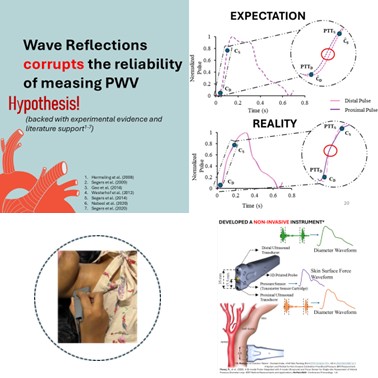
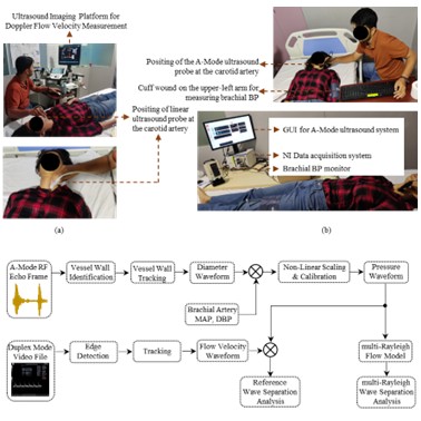
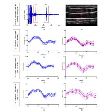
Abstract The local pulse wave velocity (PWV) from large elastic arteries and its pressure-dependent changes within a cardiac cycle are potential biomarkers for cardiovascular risk stratification. However, pulse wave reflections can impair the accuracy of local PWV measurements. We propose a method to measure pressure-dependent variations in local PWV while minimizing the influence of pulse wave reflections. The PWV is computed from the pulse transit time between two forward-traveling pulse waveforms obtained across known path length, after measured/modelled flow-based wave separation analysis (WSA). An in-vivo study of 60 participants (24 female), was conducted to compare inter- and intra-cycle variations in PWV obtained from measured and forward pulse waves. For this, proximal and distal diameter waveforms from the carotid artery, along with carotid tonometry, were recorded using a custom bi-modal arterial probe. The carotid blood flow for WSA was captured with an ultrasound imaging system. The reference PWV was derived from the Bramwell-Hill equation. After WSA, the reliability of PWV measurement improved with coefficient of variation reducing from 25% to 10% near the peak of the pulse waves and matched the reference PWV with no statistically significant difference. The average PWV at foot of the pulse wave before and after WSA were comparable to the reference PWV with no statistically significant difference. The coherence of carotid pulse pressure obtained from the mean values of PWV within a cardiac cycle after WSA with that of the carotid pulse pressure from tonometry, substantiates the results obtained for reflection-free PWV. The reliability of measuring local PWV and its pressure dependent variations within a cardiac cycle is improved by combining transit-time approach with WSA.
-
 Carotid Pressure Wave Separation Analysis Using Multi-Rayleigh Flow ModelRahul Manoj, V. Raj Kiran, P. M. Nabeel, and 2 more authorsIEEE Transactions on Biomedical Engineering, May 2025
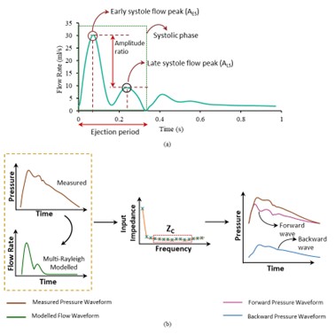
Background: Wave separation analysis (WSA) performed at the common carotid artery (CCA) reveals insights into cerebral vascular pathophysiology; however, the conventional WSA (WSAREF) necessitates the availability of both pressure and flow waveforms measured from the same arterial site. Objective: We propose a method for performing WSA at CCA using a single pulse waveform (WSAm-RAY), by modelling the shape of the CCA flow using multi-Rayleigh curves. The WSAm-RAY reduces the measurement complexity, by modelling the flow waveform shape, targeted at scenarios with limited resources, where comprehensive equipment, specialized personnel and hospital settings are often lacking. The modelled flow was compared with the measured flow for accuracy in modelling the flow morphology. The performance of WSAm-RAY was evaluated by comparing the reflection quantification indices derived from WSAREF and WSAm-RAY. Methods: WSAm-RAY employs weighted and shifted multi-Rayleigh functions, time-optimized for characteristic flow peaks in the early and late systolic phase of the CCA flow waveform. The reliability of the modelled flow and performance of WSAm-RAY were validated on 70 (28 female) participants (age: 20 to 51 years). Continuous recording of CCA flow velocity and diameter waveforms were recorded from the participants. Results: The root-mean-squared-error in forward and backward pressure waves was 2.18 ± 0.97 mmHg (∼ 2.5% of the average mean arterial pressure of the study participants). A statistically significant and strong correlation (r > 0.76, p < 0.001) was observed among reflection quantification indices from WSAREF and WSAm-RAY. Conclusion: WSAm-RAY potentially expands the scope of vascular screenings using a single pulse measurement targeting resource constrained settings.
-
 Single-element ultrasound system for high-resolution jugular venous pulse contour detectionNavya Rose George, P. M. Nabeel, Kiran V. Raj, and 3 more authorsScientific Reports, Apr 2025
Jugular venous pulse (JVP) waveform contour is directly linked to right atrial hemodynamics and is a potential tool for diagnosing various cardiovascular diseases. The reference standard, central venous line catheterization, while precise, is restricted to critical care units owing to the necessity for surgical procedures and specialized operator skills. Conventional ultrasound imaging systems can reliably measure jugular venous vessel dynamics but are hindered by low operational frame rates, large form factors, and the requirement for trained operators. In this study, we propose a portable system using single-element ultrasound technology for reliable JVP waveform acquisition and methods for quantifying the JVP contour features. The performance of the system and method was validated against a reference ultrasound imaging system in a preclinical study on 65 asymptomatic volunteers (27 female). Operating at an acquisition rate of 250 Hz, the system reliably captures JVP waveforms with a temporal resolution of 4 ms. The maximum and minimum jugular venous diameter measurements showed a statistically significant and strong correlation with the reference measurements (r = 0.93 and r = 0.86, respectively, p < 0.001). The devised algorithms effectively segmented JVP cycles and analyzed their contour features with a sensitivity and specificity of 92%. These results provide preliminary evidence for the potential use of the developed system for high-fidelity JVP waveform acquisition and pulse contour feature assessment. The ability to accurately evaluate the JVP contour characteristics can provide insights into right atrial hemodynamics, potentially facilitating the early detection and monitoring of vascular anomalies.
2024
-
 Quantification of reflected wave magnitude and transit time using a multi-Rayleigh flow waveform model: A simplified approach to arterial wave separation analysisRahul Manoj, P.M. Nabeel, V. Raj Kiran, and 2 more authorsBiomedical Signal Processing and Control, Jul 2024
Quantification of wave reflection necessitates the simultaneous measurement of pressure and flow waveforms from the same arterial site, enabling wave separation analysis (WSA). A simplified approach to WSA, which involves modelling the flow waveform, offers a methodological advantage over the conventional method. Nevertheless, the current methods of pressure-only WSA is constrained to the aortic site. In this study, we propose a method to model the carotid artery flow rate waveform using multi-Rayleigh functions (Qm-RAY(t)) for WSA (WSAm-RAY). The carotid artery is associated with central and cerebral haemodynamics and has an anatomical advantage for easier accessibility of non-invasive measurements, unlike the aorta. The accuracy of the Qm-RAY(t) was assessed against the actual carotid flow rate waveform (QREF(t)), and the performance of the WSAm-RAY was evaluated against the QREF(t) based WSA (WSAREF) on 4374 virtual (healthy) subjects. The QmRAY(t) has captured the time instants of characteristic peaks in the flow profile of the common carotid artery with an RMSE \textless 8.26 ms (\textless1 % of average heart rate). A strong and statistically significant correlation (r = 0.99, p \textless 0.001) was observed for both ΔPF and ΔPB when compared between WSAREF and WSAm-RAY. The reflection quantification indices obtained from WSAREF, and WSAm-RAY yielded strong and statistically significant corre lation (r \textgreater 0.78, p \textless 0.001). The WSAm-RAY demonstrated the capability to ease the carotid WSA by modelling the flow rate waveform and thereby expanding the possibilities for vascular screenings and diagnostics that rely on single pulse waveform measurements.
- A Feasibility Study on the Response of Vascular Impedance to Reactive HyperemiaNimmi Sudarsan, Rahul Manoj, Raj Kiran V, and 3 more authorsIn 2024 46th Annual International Conference of the IEEE Engineering in Medicine and Biology Society (EMBC), Jul 2024
Endothelium-dependent vascular reactivity has been recommended as a reliable indicator of vessel health. Ultrasound-based assessment of diameter response to shear stress measured as the proportion of brachial dilatation caused by reactive hyperemia (RH), is the gold standard for evaluating endothelial reactivity. However, the conventional approach does not account for the transient increase in blood flow and subsequent change in the transmural pressure that stimulates the endothelial cells to dilate the artery. In this work, simultaneous measures of blood flow and blood pressure during RH are used to evaluate the response of the brachial artery impedance. Endothelial reactivity is assessed using a threeelement impedance blood flow model of the brachial artery. A significant change (\textgreater 30%) was observed in the impedance parameters in response to RH. The results were repeatable (ICC \textgreater 0.64) over 2 consecutive days for 10 participants. The results demonstrate the applicability of brachial impedance analysis in evaluating endothelial reactivity. This method also directly evaluates endothelial response to shear since the flow velocity and pressure increase precede the diameter change.
- Characterizing the Effect of Hold-Down Pressure for Local and Regional Stiffness MarkersSmit Shah, Jaganathan G, Rahul Manoj, and 3 more authorsIn 2024 46th Annual International Conference of the IEEE Engineering in Medicine and Biology Society (EMBC), Jul 2024
Vascular aging occurs due to pathologies and old age. Early detection of stiffness markers and arterial geometry parameters in the carotid artery serves as an essential indicator for the progression of vascular aging. The assessment of stiffness markers is typically conducted using ultrasound and tonometerbased devices. However, both types of devices present challenges, requiring skilled operators and exhibiting variability in results due to different hold-down pressures when different operators are involved. In this study, we aim to quantify the impact of holddown pressure on diameter-based measurements and evaluate its effects on local and regional stiffness markers. To carry out this investigation, we utilized ARTSENS Plus device along with a multi-modal probe comprising an ultrasound transducer and a tonometer. A-mode ultrasound scanning was performed on the left common carotid artery of each participant, tonometer indicated the applied hold-down pressure on the participant’s skin. Four trials were conducted at hold-down pressure levels of 50, 100, 150, and 200 mmHg, and RF echo frames were recorded. ARTSENS Plus signal processing algorithms were applied to obtain the recorded frames’ carotid diameter and pressure, central pressure, and local and regional stiffness. The beat-tobeat repeatability of diameter values was examined, and the coefficient of variation was calculated to assess the consistency of the measurements. The system’s signal-to-noise ratio exceeded 25 dB. The results section delves into the impact of hold-down pressure on diameter, carotid pressure, and stiffness markers, providing insights into the variables influencing the reliability of the measurements in this cardiovascular assessment. From all the results and observations optimal hold-down pressure can be slightly higher than diastolic pressure.
- Jugular Venous Pulse Waveform Acquisition using Contact Piezo Sensor: A Pilot StudyNavya Rose George, Nimmi Sudarsan, Rahul Manoj, and 4 more authorsIn 2024 46th Annual International Conference of the IEEE Engineering in Medicine and Biology Society (EMBC), Jul 2024
The jugular venous pulse (JVP) is an important signal for detecting cardiovascular abnormalities pertaining to the right atrium. The gold standard technique of central venous line catheterization is invasive, risky, and demands expertise and hence performed only in critical care settings. Non-invasive approaches such as ultrasound and photoplethysmography are used for JVP measurement but are limited by usability issues related to the operator’s expertise. In this pilot study, we demonstrate the feasibility of acquiring JVP signals from the jugular veins (JV) using a contact piezoelectric sensor. The JVP piezo signals were acquired from 20 healthy participants and were validated against the reference ultrasound approach. The piezoelectric system could capture high-fidelity JVP signals at a resolution of 0.25 ms. The developed cycle segmentation algorithm was implemented to extract individual JVP cycles. The JVP cycles captured using the proposed and the reference methodologies yielded a correlation of 0.90 and a root mean square error (RMSE) less than 0.35. The developed pulse contour analysis algorithm evaluated the beat-to-beat JVP fiduciary marker locations with a maximum coefficient of variation of 17 %. The piezoelectric sensor measurements were more susceptible to motion artifacts in comparison to the reference ultrasound system. To our knowledge, this is the first use of contact piezo sensors for direct measurement of JVP signals. Future advancements based on this technique can provide viable options for ambulatory and self-measurementbased cardiac health screening.
- Wearable Acceleration Plethysmography for Carotid Pulse Pressure Monitoring: A Feasibility StudyV V Girish, Rahul Manoj, P. M. Nabeel, and 1 more authorIn 2024 46th Annual International Conference of the IEEE Engineering in Medicine and Biology Society (EMBC), Jul 2024
Central blood pressure (CBP), which is the pressure at the aorta and other central arterial sites such as the carotid, is more predictive of target organ damage and future adverse events, as opposed to conventional blood pressure assessed at more peripheral sites. However, methods to estimate CBP are complex, require frequent calibration and are not suitable for continuous tracking purposes. Here, we introduce a wearable accelerometer based pressure sensing system to track carotid pulse pressure (PP). The wearable system detects the transcutaneous pressure (PT) on the skin surface over the carotid artery and identifies the key fiducial points (g1, g2). In-vivo study on 11 subjects showed that the peak-to-peak value, ΔPT and maximum value, g2 of PT have a statistically significant and strong correlation with carotid PP (r = 0.75, p \textless 0.05) and SBP (r = 0.78, p \textless 0.05) respectively. The magnitude response (percentage mean difference between pre and post-exercise intervention) of PT parameters (ΔPT and g2) were 78 % and 89 % as compared to 33 % and 19 % in carotid PP and systolic blood pressure (SBP), respectively. Reliability (SNR \textgreater 38 dB) and repeatability (CoV \textless 10%) were seen in the obtained transcutaneous pressure signals. The study limitation is identifying an appropriate calibration or model for carotid BP estimation. This study reveals the feasibility of using an acceleration plethysmogram (APG) based wearable pressuresensing system for tracking changes in carotid PP and SBP which could pave the way for wearable systems for ambulatory assessment of central BP for clinical and home use.
- Acoustic Plethysmography for Aortic Pulse Wave Velocity Measurement: In-Vitro and In-Vivo Feasibility StudyIshwarya S, Raj Kiran V, Rahul Manoj, and 2 more authorsIn 2024 IEEE International Symposium on Medical Measurements and Applications (MeMeA), Jun 2024
Large arterial stiffness is an indicator of vascular aging and is a prognostic marker of cardiovascular disease. Carotid-femoral pulse wave velocity (cf-PWV) is a gold standard technique for non-invasively assessing large artery stiffness. The state-of-the-art cf-PWV measuring devices necessitate expertise and are not pertinent for routine care in non-clinical settings. To address this gap, we have developed an acoustic plethysmography device for cf-PWV measurement. An in-vitro test on an arterial phantom was performed to ensure the device’s reliability. Subsequently, an in-vivo study of 10 participants was conducted to assess the feasibility of the acquisition of the developed device. The in-vitro measurement accuracy was satisfactory, with a root mean square error of .4%. The in-vivo PWV results are consistent, with the normal ranges for the measured age group falling between 4 and 5.8 m/s. The beat-to-beat COV obtained from in-vitro and in-vivo studies were less than 4.2% and 4.86% respectively. The study’s findings demonstrated that the proposed device has the potential to seamlessly provide cf-PWV. However, additional validation against the reference devices on a larger population is in progress and will be published in future publications.
- Assessment of Local Venous Pulse Wave Velocity Using Single-Site Methods- A Pilot StudyNavya Rose George, Rahul Manoj, Raj Kiran V, and 3 more authorsIn 2024 IEEE International Symposium on Medical Measurements and Applications (MeMeA), Jun 2024
There has been a growing interest in assessing pulse wave velocity (PWV) in the veins with studies highlighting their potential in elucidating the vascular indices such as the venous pressure changes, venous return dynamics, venous insufficiency, and volaemic status. Despite their clinical significance, the measurements are limited due to the challenges associated with reliable venous pressure assessments. The local venous pulse wave velocity (vPWV) refers to the speed of pulse propagation within a targeted vein. Conventionally, PWV is calculated using a pulse-transit time-based approach where the venous pulses are measured from two known locations, necessitating the acquisition of high-fidelity venous pulses and demanding operator expertise. This study explores the feasibility of measuring local venous pulse wave velocity using established single-site measurement approaches employed in the arterial domain. These methods rely on simultaneous diameter and blood flow velocity signals from the vessel, employing the diameter-flow velocity (ln(D)U) and area-flow rate (QA) relationship in the vein. A pilot feasibility study was conducted on 16 participants aged 20-35 years, with the internal jugular vein selected as the target vein due to its proximity to the right atrium. Jugular venous diameter and flow velocity signals were captured at a resolution of 2 ms (frame rate = 500 Hz), and local vPWV values were derived. The in vivo measurements yielded local vPWV values, ranging from 0.5 ms-1 to 2.2 ms-1, consistent with earlier literature findings. Furthermore, both measurement approaches demonstrated a significant correlation (r = 0.95, p \textless 0.05), validating their efficacy in assessing local venous pulse wave velocity.
- Estimating Left Ventricular Contractility Through Carotid Artery Distension: A Portable Device Utilizing A-Mode Ultrasound and Surrogate Marker AnalysisGanapathy Jaganathan, Rahul Manoj, Raj Kiran V, and 2 more authorsIn 2024 IEEE International Symposium on Medical Measurements and Applications (MeMeA), Jun 2024
Non-invasive assessment of left ventricular (LV) function is crucial for early diagnosis and management of cardiovascular diseases. The Acceleration Time (AT) to Ejection Time (ET) ratio, is a measurement used to assess the contractility of the left ventricle (LV) of the heart. This study explores the feasibility of a novel device for a surrogate estimation of the AT/ET ratio using carotid artery distension dynamics. In this in-vivo study, 17 healthy young male participants were recruited. A custom-designed device combining A-mode ultrasound and a pocket Doppler was used to acquire simultaneous measurements of carotid artery distension and blood flow velocity. The pocket Doppler aided in identifying critical events in the carotid artery flow waveform, serving as reference points for analyzing the second derivative of the carotid distension waveform obtained by the A-mode component. This facilitated the estimation of surrogate AT, ET, and the AT/ET ratio from carotid artery dynamics. The study demonstrated a strong correlation between surrogate markers and reference measurements. The surrogate AT measured from carotid distension showed a significant correlation (r = 0.81, p \textless 0.05) with the reference AT obtained from LVOT flow. Similarly, the surrogate ET derived from carotid distension exhibited a strong correlation (r = 0.83, p \textless 0.05) with the reference ET from LVOT. Importantly, the surrogate AT/ET ratio estimated from carotid artery dynamics displayed a significant correlation (r = 0.8, p \textless 0.05) with the reference AT/ET ratio measured using the gold-standard LVOT flow waveform. This study suggests that the combined A-mode ultrasound and pocket Doppler device has the potential to serve as a non-invasive tool for estimating key LV function parameters, including the clinically relevant AT/ET ratio, through analysis of carotid artery distension. Further research with a larger and more diverse population is warranted to validate this approach for broader clinical applications.
- Impact of Physical (In)Activity on Carotid-Femoral PWV and Central Blood Pressure in Young and Middle-Aged Adults: A Pilot Study using ARTSENS PlusGanapathy Jaganathan, Rahul Manoj, Raj Kiran V, and 2 more authorsIn 2024 IEEE International Symposium on Medical Measurements and Applications (MeMeA), Jun 2024
Carotid-femoral pulse wave velocity (cfPWV) and central blood pressure (BP) are prognostic stiffness markers for assessing vascular health. In this study, a group of young (20 ≤ age \textless 40) to middle-aged (40 ≤ age \textless 60) participants with minimal cardiovascular risk were examined to determine the impact of physical (in)activity on central BP and cfPWV. This in-vivo study was conducted on 97 healthy participants (Mean age: 34) . All participants completed a detailed lifestyle and medical history questionnaire, which included information on their lifestyle and physical activity. An in-house developed A-mode ultrasound device, ARTSENS Plus was used to measure, central BP (from the carotid artery) and cfPWV. There was no significant difference in central BP and cfPWV between physically active and inactive young adults (p \textgreater 0.05). Although cfPWV in middle-aged adults increased by 26% in females and 10% in males between active and inactive populations, the observation was not statistically significant (p = 0.14 in males and p = 0.11 in females). The difference in carotid pulse pressure between physically active and inactive middleaged adults was statistically significant (p ≤ 0.05). Results from this study will eventually cast new light on the importance of physical (in)activity among young and middle-aged populations.
-
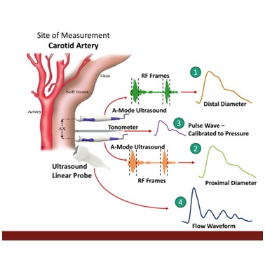 Measurement of Inter and Intra-cycle Variations in Local Pulse Wave Velocity from Forward Travelling Pulse WavesRahul Manoj, Raj Kiran V, Nabeel P M, and 2 more authorsIn 2024 IEEE International Symposium on Medical Measurements and Applications (MeMeA), Jun 2024
The local PWV and its pressure-dependent changes within a cardiac cycle are potential biomarkers for cardiovascular risk stratification. However, the influence of pulse wave reflections hampers the accuracy of local PWV measurements. We propose a method to measure the pressuredependent variations in local PWV after minimizing the effect of wave reflections. The technique measures the pulse transit time between two simultaneously acquired and processed forward-running pulse waveforms obtained after wave separation analysis (WSA). A cross-sectional in-vivo study was conducted on 57 human participants (22 female) to compare the inter and intra-cycle variations in PWV based on pulse transittime obtained from two measured pulse waves and two forwardtravelling pulse waves. The reference PWV for comparison was obtained using Bramwell-Hill equations. The required signals –proximal and distal diameter waveforms from the carotid artery, simultaneously with carotid tonometry were acquired using an in-house developed bi-modal arterial probe. The carotid blood flow for WSA was recorded using an ultrasound imaging system. The reliability of measuring PWV was improved after WSA (coefficient of variation improved from 25% to 10% for PWV at the systolic level). The average PWV obtained before WSA and after WSA at diastolic levels were comparable with the reference PWV (p \textgreater 0.05). The average PWV obtained before WSA and after WSA at systolic levels were significantly different (p \textless 0.001); however, the PWV after WSA at systole level was comparable with the reference PWV (p \textgreater 0.05). The group average carotid pulse pressure derived from the mean values of PWV within the cardiac cycle, obtained after WSA (25.90 ± 5.88 mmHg), was statistically (p \textgreater 0.05) comparable to the carotid pulse pressure obtained using carotid tonometry-calibrated with brachial mean and diastolic blood pressure (26.74 ± 4.72 mmHg). Therefore, this proposed method has the potential to improve the measurement reliability of local PWV, aiding the ongoing global efforts of cardiovascular risk management.
-
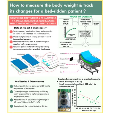 Periodic Weight Measurement for Bedridden Patients Using a Pressurized Liquid-Filled Channel System Integrated with Hospital BedsRahul Manoj, Ramdayalan Kumarasami, Gunjan Singh, and 3 more authorsIn 2024 IEEE International Symposium on Medical Measurements and Applications (MeMeA), Jun 2024
Variations in body fluids in a short time can lead to significant health issues. The risk due to body fluid variations increases for critically ill and bedridden patients. Changes in body weight in short intervals are the primary indicator of body fluid imbalances. The clinical practice for periodic monitoring of body weights of critically ill or bedridden patients includes a dedicated weighing scale or lift scale, detachable or built-in units of weighing scale on to the legs of hospital cots or as integrated bed scales attached permanently to the cot. However, these methods have limited mobility and usability in a practical setting and often require the involvement of multiple personnel to measure the body weight of a bedridden patient. Owing to such constraints, periodic monitoring of body weights is seldom recorded. In this work, we propose a novel measurement scheme for periodic monitoring of body weight. The prototype consists of an active (piston-based syringe pump) pressurized liquidfilled flexible and elastic channel sandwiched between the hospital cot and mattress. The required displacement of the syringe pump to achieve an optimal internal pressure is calibrated with the known weights. A calibration model (R2 = 0.99, p \textless 0.001) is developed that maps the displacement of the piston pump in mm to known weights in kg for the range 0 to 90 kg. Using the calibration model, the accuracy and precision of the system are characterized at various loading conditions. A practical scenario of rising body weights was emulated as an invitro-experiment, resulting in a maximum error of 3%. The design of the system is modular, so it can easily be integrated with any type of hospital bed. This design is proof of concept for using alternative technologies for weight measurement over periodic intervals, where conventional designs seem cumbersome.
2023
- A Pilot Observational Cohort Study to Investigate the Effect of Valsalva Maneuver on Internal Jugular Venous DiameterNavya Rose George, Rahul Manoj, Raj Kiran V, and 4 more authorsIn 2023 45th Annual International Conference of the IEEE Engineering in Medicine & Biology Society (EMBC), Jul 2023
Valsalva maneuver (VM) is a technique widely used for acute elevation of blood pressure in humans. It has potential applications in cardiac health prediction and is also a diagnostic tool in cardiovascular, neurology and ENT screening. The jugular venous (JV) diameter increases during the VM procedure and hence it has been widely used to aid central venous catheterization in medical units. In this pilot study, we have quantified the variation in JV diameter response to VM across young and middle-aged populations. The study was conducted on a cohort of 16 males and 11 females, where the JV diameter in baseline, during and post VM intervention were acquired using a B-mode imaging system. The JV diameter measurements were within the ranges specified in earlier literature. The beat-to-beat variability in baseline diameter measurements was found to be between 8% to 20%. In younger population, the average maximum JV diameter during baseline was found to be 9.25 ± 2.61 mm and in middle-aged population it was 12.49 ± 2.65 mm. The average maximum JV diameter in young and middle-aged population during VM was 11.66 ± 2.74 mm and 16.73 ± 3.28 mm respectively. The study findings suggested a statistically significant variation (p \textless 0.05) between the JV diameter responses from young and middle-aged populations. The JV distensibility decreased significantly during VM in younger cohort (-35%) in comparison with the minimal changes observed in middle-aged population. The study demonstrates the variation in JV diameter and distensibility to VM in young and middle-aged populations.
- Arterial Wave Separation Analysis and Reflection Wave Transit Time Estimation using a Double Rayleigh Flow Rate ModelRahul Manoj, Aneesh S, Raj Kiran V, and 3 more authorsIn 2023 45th Annual International Conference of the IEEE Engineering in Medicine & Biology Society (EMBC), Jul 2023
Arterial pulse wave separation analysis (WSA) requires simultaneously measured pressure and flow rate waveform from the same arterial site. Modelling approaches to flow rate waveforms offers a methodological and instrumentational advantage. However, current techniques are limited to the aortic site. For non-aortic sites such as carotid artery, modelling methods that were developed for aortic sites are not likely to capture the intrinsic differences in the carotid flow rate. In this work, a double-Rayleigh flow rate model for the carotid artery is developed to separate the forward and backward pressure waves using WSA (DRMWSA). The model parameters are optimally found based on characteristic features - obtained from the pressure waveform. The DRMWSA was validated using a database of 4374 virtual (healthy) subjects, and its performance was compared with actual flow rate based WSA (REFWSA) at the carotid artery. An RMSE \textless 2 mmHg were obtained for forward and backward pressure waveforms. The reflection quantification indices (ΔPF, ΔPB), (RM, RI) obtained from DRMWSA demonstrated strong and statistically significant correlation (r \textgreater 0.96, p \textless 0.001) and (r \textgreater 0.80, p \textless 0.001) respectively, with insignificant bias (p \textgreater 0.05), upon comparing with counterparts in REFWSA. A moderate correlation (r = 0.64, p \textless 0.001) was obtained for reflection wave transit time between both methods. The proposed method minimises the measurements required for WSA and has the potential to widen the vascular screening procedures incorporating carotid pulse wave dynamics.
- Assessment of Endothelial Reactivity by Measurement of Vascular Material Response to Shear Stress: A Feasibility Study *Nimmi Sudarsan, Rahul Manoj, Raj Kiran V, and 3 more authorsIn 2023 45th Annual International Conference of the IEEE Engineering in Medicine & Biology Society (EMBC), Jul 2023
Flow-mediated dilation (FMD) evaluates the relative change in arterial diameter during hyperemia to assess the endothelial response due to a shear stimulus. However, conventional FMD measures diameter response alone and the alterations in the arterial wall’s material properties during reactive hyperemia, which also influence dilation, go unaddressed. In this work, we examine the material response (MR) of the artery during reactive hyperemia using clinically relevant stiffness markers for the assessment of endothelial reactivity (ER). For this, we have developed an in-house brachial cuff control (BCC) system to continuously acquire brachial pressure which can be integrated with simultaneous measurement of brachial diameter and used to quantify the relative changes in wall property during hyperemia noninvasively. The assessment of endothelial reactivity using material response (ERAMR) was conducted on 20 healthy participants (12M/8F) and the results were compared with conventional FMD (FMD%). The mean pressure response gave an inverse trend to that of diameter response with varying magnitudes during reactive hyperemia (18.71% from baseline for diameter and 2.45% for pressure), there was a significant difference in the measurement of FMD and ERAMR (P \textless 0.05). The larger distribution of ERAMR compared to FMD% in boxplots further implies the inclusion of within-subject variations. Hence, ERAMR can be a potential estimate of ER, given the need for intensive validations in this line on larger cohorts.
- Normalization of Flow-mediated Dilation to Brachial Artery Material Property: A Feasibility Study^\textrm*Nimmi Sudarsan, Rahul Manoj, Raj Kiran V, and 3 more authorsIn 2023 45th Annual International Conference of the IEEE Engineering in Medicine & Biology Society (EMBC), Jul 2023
Endothelial reactivity (ER) is widely measured using flow-mediated dilation (FMD) of brachial artery. Conventional measurement of FMD is influenced by factors such as input shear stress, arterial transmural pressure, diameter and thereby arterial material properties (ℇ). Thus, for a reliable interpretation of FMD, it has to be normalized with respect to the above confounding factors. Normalization of FMD with shear stress at the time of measurement has been reported to reduce measurement variability. However, its widespread usage among the research community is limited. In this work, we examine the feasibility of normalizing the brachial FMD index (FMD%) to ℇ : extrema (ℇ ), baseline (ℇ ) and extrema change (∆ℇ) post-ischemia using its inter-day variability against FMD. In-vivo measurements were performed on 10 participants for 2 consecutive days and simultaneous pressure-diameter cycles were collected to estimate the material properties during reactive hyperemia (RH). The box-whisker plot reveals differences in the mean and deviation of FMD to FMD\textbarℇb. A significant value for repeatability (ICC ≥ 0.6) was obtained for normalized FMD (FMD\textbarℇb) for specific stiffness index (β), pressure-strain elastic modulus (Ep), and local pulse wave velocity (PWV) as compared to FMD. Hence, normalization of FMD% to arterial ℇ can potentially improve the measurement reliability of ER assessment.
- Ultrasound for Venous Local Pulse Wave Velocity: Comparison of Pulse Transit Time MethodsNavya Rose George, Rahul Manoj, Raj Kiran V, and 3 more authorsIn 2023 45th Annual International Conference of the IEEE Engineering in Medicine & Biology Society (EMBC), Jul 2023
Venous pulse wave velocity (vPWV) is a potential marker for determining the state of venous hemodynamics, venosclerosis, and vascular filling. Although there have been several studies on pulse wave velocity through blood vessels, the majority have focused on arteries, with only limited studies on veins. To our knowledge, this study is the first to compare the local vPWV estimation metrices. An in vivo study was conducted on 10 participants where the jugular venous pulses (JVP) from two proximal sites were simultaneously acquired using a dualelement high frame rate system. The local vPWV was computed using different transit time-based techniques. The study demonstrates the comparison between vPWV ranges computed using thresholding, fiduciary point (c and v) and correlationbased approaches indicated as vPWV\textbarth, vPWV\textbarc, vPWV\textbarv and vPWV\textbarXcorr respectively. High fidelity echo frames were acquired from the jugular vein (JV) at a temporal resolution of 2 ms and an amplitude resolution of 10 µm. The study findings indicated that the vPWV computed using various transit time metrics were comparable without significant bias (p \textgreater 0.05). Among the VPWV metrices, vPWV\textbarth had the lowest beat-to-beat variation (CoV = 18 %). The mean deviations in vPWV\textbarc, vPWV\textbarv and vPWV\textbarXcorr values from vPWV\textbarth were 0.28, 0.17 and 0.22 m/s respectively, where the average beat-to-beat variation was minimal. The results suggested that the thresholding and crosscorrelation metrices offered better performance in comparison with the fiduciary point techniques for vPWV estimation.
-
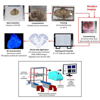 Wide field block face imaging using deep ultraviolet induced autofluorescence of the human brainSrinivasa Karthik, Jayaraj Joseph, Jaikishan Jayakumar, and 6 more authorsJournal of Neuroscience Methods, Sep 2023
Background: Imaging large volume human brains at cellular resolution involve histological methods that cause structural changes. A reference point prior to sectioning is needed to quantify these changes and is achieved by serial block face imaging (BFI) methods that have been applied to small volume tissue (~1 cm3). New method: We have developed a BFI uniquely designed for large volume tissues (~1300 cm3) with a very large field of view (20 × 20 cm) at a resolution of 70 µm/pixel under deep ultraviolet (UV-C) illumination which highlights key features. Results: The UV-C imaging ensures high contrast imaging of the brain tissue and highlights salient features of the brain. The system is designed to provide uniform and stable illumination across the entire surface area of the tissue and to work at low temperatures, which are required during cryosectioning. Most importantly, it has been designed to maintain its optical focus over the large depth of tissue and over long periods of time, without readjustments. The BFI was installed within a cryomacrotome, and was used to image a large cryoblock of an adult human cerebellum and brainstem (~6 cm depth resulting in 2995 serial images) with precise optical focus and no loss during continuous serial acquisition. Comparison with existing method(s): The deep UV-C induced BFI highlights several large fibre tracts within the brain including the cerebellar peduncles, and the corticospinal tract providing important advantage over white light BFI. Conclusions: The 3D reconstructed serial BFI images can assist in the registration and alignment of the micro scopic high-resolution histological tissue sections.
- Assessment of Endothelial Reactivity using Brachial Pulse Wave Velocity Response to ShearNimmi Sudarsan, Rahul Manoj, V Raj Kiran, and 3 more authorsIn 2023 IEEE International Symposium on Medical Measurements and Applications (MeMeA), Jun 2023
Non-invasive assessment of endothelial reactivity (ER) is done by analyzing the relative change in brachial artery diameter in response to transient forearm ischemia. However, confounding factors such as flow velocity that mediates to dilation of the artery are not considered while calculating the ER index (FMD%). Therefore, we propose a technique for the assessment of ER using the measurement of local pulse wave velocity (PWV) during reactive hyperemia (RH). Continuous diameter and flow velocity were recorded on 10 healthy subjects during baseline, RH and recovery phases and corresponding PWV was calculated using the flow-area (QA) and ln(diameter)velocity (DU) method. After 54 ± 32s of cuff release, PWV increased to 35.53% and 18.45% compared to baseline, and recovered to 45.6% and 55.62% from RH in 78 ± 16s for both QA and DU methods. The study was repeated for 2 consecutive days. Repeatable diameter and flow measurements were obtained (ICC \textgreater 0.6). There existed a significant mean difference in estimation of PWV using QA and DU methods (P \textless 0.05). The ICC for PWV derived ER was 30% higher than FMD%. Hence, analyzing temporal response of local PWV to ischemia is a reliable technique for assessment of ER. Direct methods for evaluating PWV during the course of the experiment can be a potential and simpler technique for assessment of ER.
- Effect of Fiduciary Point Choice on Pulse Wave Velocity-based Cuffless Pulse Pressure Estimation: Ex-vivo StudyRaj Kiran V, Rahul Manoj, S Ponkalaivani, and 2 more authorsIn 2023 IEEE International Symposium on Medical Measurements and Applications (MeMeA), Jun 2023
Superiority of central blood pressure (BP) (especially its pulsatile component; pulse pressure (PP)) over that of brachial has been underlined recently through several clinical studies. Local pulse wave velocity (PWV) based cuffless methods using Bramwell-Hill (BH) equation are popularly employed to assess PP. These approaches assume PWV to be constant, whereas, it changes with pressure. Pertaining to this, literature is unclear on which instantaneous local PWV value should be chosen within the cardiac cycle for PP evaluation. Since local PWV can be measured from various fiducial points spread across the blood pulse cycle, it may be relevant to investigate on the choice of particular one(s) for reliable PP calculation. We have conducted an ex-vivo study in this regard employing an excised ovine artery, emulating 21 independent flow conditions by changing the PP and mean arterial pressure (MAP). The measured end-diastolic (ED) PWV was lower than the peaksystolic (PS) PWV by 32%, and in theory they are the extremities of PWV within a cardiac cycle. An underestimation of 26% was observed in the PP evaluated using ED-PWV and overestimation of 30% using PS-PWV. The PWV that is expected to yield an exact PP value corresponded to the instant in the blood pulse cycle where its mean occurred. The ED-PWV underestimated and the PS-PWV over-estimated the expected PWV by 17% and 14%, respectively, which explains the deviations in the estimated PPs. The time instant of the first derivate maximum being closer to that of the cycle’s mean makes it a potential choice for measuring PWV for PP estimation.
- Measurement of Local Pulse Wave Velocity: Agreement Among Various MethodologiesRahul Manoj, V Raj Kiran, S Ponkalaivani, and 3 more authorsIn 2023 IEEE International Symposium on Medical Measurements and Applications (MeMeA), Jun 2023
The local pulse wave velocity (PWV) is the velocity with which the arterial pulse wave travels from the left ventricle to the vascular bed. Local PWV is clinically significant as a prognostic indicator of vascular damage. The measurement of local PWV involves several direct and indirect methods. However, there are limited studies that compare agreement among different methodologies. In this work, we investigated the agreement among several methods of measurement of PWV, such as the haemodynamic loop-based, Bramwell-Hill, transittime-based and computational models of PWV. A small cohort of 35 participants (21 male/14 female) aged between 21 and 51 years was recruited on voluntary consent. The measurement setup included duplex mode recording of carotid diameter and flow velocity waveforms from an ultrasound machine and simultaneous acquisition of dual-diameter waveforms and tonometry waveforms using an in-house developed bi-modal arterial probe. The carotid pressure waveform, flow velocity and dual diameter waveforms for evaluating the various methods of PWV measurement were obtained from the data processing. The group average value of PWV were obtained between 3.07±1.17 m/s to 5.02±1.00 m/s for various methods. The lowest and the highest group average PWV was reported using the haemodynamics-loop-based methods. There exists a strong and statistically significant correlation among PWV obtained using Bramwell-Hill equations and computational models (r \textgreater 0.91, p \textless 0.001), whereas a moderate and statistically significant correlation was observed between Bramwell-Hill and transit-time-based methods (r = 0.67, p \textless 0.001). The correlation was poor between Bramwell-Hill and loop-based methods (r ~ 0.2, p \textless 0.001). The study confirms the variations in the measurements in PWV using different methods and suggests their interchangeable usage is not advised.
2022
- Association of Local Arterial Stiffness and Windkessel Model Parameters with Ageing in Normotensives and HypertensivesNimmi Sudarsan, Rahul Manoj, Nabeel P M, and 2 more authorsIn 2022 44th Annual International Conference of the IEEE Engineering in Medicine & Biology Society (EMBC), Jul 2022
Computation of arterial stiffness is a well-established, widely accepted method for estimating vascular age. Although carotid-femoral pulse wave velocity is typically used for vascular age assessment, most recent studies have reported the need to consider a combination of local and regional stiffness indices possessing distinct association with the vascular structure and/or function for better prediction of early vascular ageing syndrome. In this work, we investigate the association of clinically validated local stiffness (obtained using biomechanical relations), global stiffness (obtained from 3-element Windkessel modelling), and pulse contour indices from the aorta with ageing and their distribution in normotensives and hypertensives. The analysis was performed on 420 (virtual) subjects (age: 65 ± 11 years) with an equal proportion of hypertensive (age: 65 ± 11 years) and normotensive (age: 65 ± 11 years) subjects. Multivariate linear regression analysis revealed an independent association of each of the indices with age (Adjusted r = 0.75 p \textless 0.01). Specific stiffness index (r = 0.67, p \textless 0.001), Augmentation index (r = 0.55, p \textless 0.001) and total arterial compliance (r = -0.50, p \textless 0.001) depicted highest correlation with age. There was a significant difference (\textgreater 16%, p \textless 0.001) in mean values of the measured indices between hypertensive and normotensive subjects. The study findings further emphasize the need to combine multiple non-invasive vascular markers to capture the unique aspects of age-induced arterial wall remodelling for reliable monitoring and management of the early vascular ageing syndrome.
- Comparison of Approximated and Actual Bramwell-Hill Equation Implementation for Local Pulse Wave Velocity: Ex-vivo StudyRaj Kiran V, Rahul Manoj, Ishwarya S, and 2 more authorsIn 2022 44th Annual International Conference of the IEEE Engineering in Medicine & Biology Society (EMBC), Jul 2022
Bramwell-Hill (BH) equation is widely adopted for the evaluation of local pulse wave velocity (PWV), primarily for its theoretical association with the vessel’s distensibility. Its implementation, however, requires arterial pressure and diameter waveforms simultaneously from a single site. Owing to the challenges associated with such a noninvasive recording, an approximated BH equation is adopted without requiring the entire pressure waveform but only the diastolic and systolic values. The approximated BH method yields a single value of local PWV as opposed to the actual method that provides instantaneous PWV within a cardiac cycle. This study aims to provide the currently lacking insights into how the approximate versus actual BH implementations compare. The study also addresses the pivotal question of which instantaneous value within the cardiac cycle corresponds to the approximated BH. An ex-vivo study was conducted for this purpose, emulating different flow conditions (changing mean and pulse pressures) to vary the local PWV within the range of 4.4 to 8.9 m/s. The results revealed the expected (pressure-dependent) incremental nature of local PWV due to hyper-elastic behavior of the artery, with systolic BH-PWV \textgreater diastolic BH-PWV by 13.6%. The approximate BH-PWV was similar to actual BH-PWV obtained from mean pressure level. It further underestimated the systolic, and overestimated the diastolic PWVs by 8.5% and 6.6%, respectively.
- Estimation of Characteristic Impedance using Multi-Gaussian Modelled Flow Velocity Waveform: A Virtual Subjects StudyRahul Manoj, V Raj Kiran, P M Nabeel, and 2 more authorsIn 2022 44th Annual International Conference of the IEEE Engineering in Medicine & Biology Society (EMBC), Jul 2022
Characteristic impedance (ZC) of the blood vessel relates the pulsatile pressure to pulsatile blood flow velocity devoid of any wave reflections. Estimation of ZC is useful for indirect evaluation of local pulse wave velocity and crucial for solving wave separation analysis (WSA) which separates the forward-backward pressure and flow velocity waveforms. As opposed to conventional WSA, which requires simultaneous measurement of pressure and flow velocity waveform, simplified WSA relies on modelled flow velocity waveforms, mainly introduced for the aorta. This work uses a multi-Gaussian decomposition (MGD) modelled flow velocity waveform to estimate ZC by employing a frequency domain analysis, which is applicable to other arteries such as carotid. Thus obtained ZC is compared with ZC estimated from true flow velocity waveform for healthy (virtual) subjects taken for the carotid artery. The MGD modelled flow velocity waveform estimated ZC for a range of 4.98 to 34.79 with a group average of 16.43±0.10. The difference between the group average values of both ZC was only 4.72%. A statistically significant and strong correlation (r = 0.708, p \textless 0.0001) was observed for ZC obtained from MGD modelled flow velocity waveform with ZC obtained from actual flow velocity waveform. The bias for ZC between the two methods was 0.74, with confidence intervals (CIs) between 7.44 and -5.96 for the Bland-Altman analysis. Therefore, ZC from MGD modelled flow velocity waveform is a potential surrogate of the flow velocity model for WSA at the carotid artery.
- Evaluation of Pulse Contour Markers using an A-Mode Ultrasound: Association with Carotid Stiffness Markers and AgeingRahul Manoj, V Raj Kiran, Pm Nabeel, and 2 more authorsIn 2022 44th Annual International Conference of the IEEE Engineering in Medicine & Biology Society (EMBC), Jul 2022
Vascular ageing is directly associated with the blood vessel wall structural and functional abnormalities. Pulse morphology carries information on these abnormalities, and pulse contour analysis (PCA) identifies key amplitudes and timing information on the pulse waveforms that has a prognostic value towards cardiovascular risk stratification. PCA markers derived from second derivative waveforms represent the accelerative and decelerative phase of an arterial pulse. In this work, second derivative diameter waveforms of central arteries such as carotid artery are obtained using an A-mode ultrasound device. The derived PCA markers (b/a, c/a, d/a, e/a, (b-c-d-e)/a) from diameter waveform is investigated for its association with central stiffness markers and aging. An observational and crosssectional study on 106 subjects (51 male/55 females) was conducted for this investigation. The highest correlation (r = 0.5, p \textless 0.001) was observed between c/a and PWV, and the lowest correlation was between e/a and AC. Group average values of PCA markers for each age decade group were correlated strongly (r \textgreater 0.9, p \textless 0.001) with age. A change \textgreater 19% was observed between the group average values of PCA markers of the normotensive and hypertensive population. The applicability of aforesaid PCA markers on central pulse waveforms, measured using a noninvasive device in resource-limited field settings, would accelerate such large scale vascular screening that is essential to understanding the cardiovascular risks at a population level.
- Operator Variabilities in Carotid Pulse Wave Velocity Measured by an Image-free Ultrasound DeviceRaj Kiran V, Rahul Manoj, Ishwarya S, and 2 more authorsIn 2022 44th Annual International Conference of the IEEE Engineering in Medicine & Biology Society (EMBC), Jul 2022
Local pulse wave velocity (PWV) has gained much attention in the last decade due to its ability to provide localized stiffness information from a target vessel and cater to several applications beyond regional PWV. Transit time-based methods are the most straightforward, but their reliability is highly dependent on the blood pulse sensing modality. Conventional ultrasound systems directly measure the blood pulse (as diameter or flow velocity); however, they offer limited frame rates resulting in poor resolution signals. Advanced systems supporting high frame rates are expensive, complex, and not amenable to field and resource-constraint settings. We have developed a high frame image-free ultrasound system to address this gap for automated and online measurement of local PWV. In an earlier in-vitro study, we have demonstrated its accuracy. In this work, we aim to investigate its in-vivo reliability. A study on 15 young, healthy subjects was conducted to assess the intraand inter-operator repeatability of the developed system. The yielded local PWVs from the left carotid artery were within the range of 2.5 to 5.8 m/s. The device provided highly repeatable intra- and inter-operator measurements with ICC of 0.94 and 0.88, respectively. The bias for the intra- and inter-operator trials was statistically negligible (p \textgreater 0.005). The study demonstrated the potential of the high frame rate device to perform reliable measurements in-vivo.
- Assessment of Arterial Reflection Markers using an A-Mode Ultrasound DeviceRahul Manoj, V Raj Kiran, Pm Nabeel, and 2 more authorsIn 2022 IEEE International Symposium on Medical Measurements and Applications (MeMeA), Jun 2022
Reflections of arterial blood pulse waves have a pivotal role in the equilibrium of the vasculature. Elevated levels of wave reflections cause an increase in pulse pressure and pulse propagating speeds, exacerbating cardiovascular risk. Quantification of reflection markers is either based on augmentation index or reflection magnitude (RM) and reflection index (RI), both derived from wave separation analysis (WSA). Simultaneous measurement of pressure and flow velocity from the same arterial site is a requirement for WSA and has its practical challenges. Subsequently, simplified WSA based on modelling flow is proposed. This work explores the feasibility of using multi-Gaussian decomposition (MGD) of diameter scaled pressure waveform to perform a WSA and quantify the reflection markers. The diameter waveforms are obtained using an A-mode ultrasound device (ARTSENS®). The decomposed pressure signals scaled from diameter waveforms (or Gaussians) are uniquely combined to yield a forward and backward wave. The reflection markers derived from MGD based WSA are then compared with the clinically relevant stiffness markers and with age. The study was conducted on 110 healthy subjects (60 males and 50 females). A moderately significant correlation (r \textgreater 0.51, p \textless 0.001) was obtained for RM and RI when compared with stiffness markers (β, Ep, AC, PWV and AIx). The highest correlation was observed for RM versus Ep (r = 0.602, p \textless 0.001), followed by β and PWV. The correlation in reflection markers with age was captured with r = 0.51, p \textless 0.001. A change of 25.2% and 15.4% were observed for the group average RM and RI, respectively, among normotensive and hypertensive subjects in this cohort. The proposed MGD model has the potential to explore the central arterial biomechanics from a diameter or pressure waveform. The variations in reflection markers with stiffness and age derived using the proposed WSA approach were faithfully captured. The flow-independent WSA, combined with a field-deployable measurement device like ARTSENS®, has the potential to conduct large scale vascular screenings in a resourcelimited setting.
- Association of Windkessel Model Parameters with Local and Regional Aortic Stiffness IndicesNimmi Sudarsan, Rahul Manoj, Nabeel P M, and 2 more authorsIn 2022 IEEE International Symposium on Medical Measurements and Applications (MeMeA), Jun 2022
Windkessel model assesses the buffering function of large arteries and has the potential to describe the global characteristics of the vasculature. In this work, we investigate the association between the Windkessel model parameters and the existing, clinically significant local, global stiffness indices and blood pulse indices. A three-element Windkessel model was used to estimate the ventricular-arterial coupling, arterial compliance, and peripheral resistance from the pressure and flow waveforms at the aorta. The study was performed on 213 healthy virtual subjects (age: 44 ± 15 years). Total arterial compliance had a significant individual correlation (R \textgreater 0.50, p \textless 0.05) with local and regional stiffness indices whereas characteristic impedance had a correlation of (R \textgreater 0.22, p \textless 0.05) with stiffness indices. Total peripheral resistance, on the other hand, did not show a significant correlation with vascular stiffness. Multivariate regression gave a better independent correlation between the Windkessel model parameters and vascular indices (Adjusted R \textgreater 0.88, p \textless 0.05) with total peripheral resistance showing the highest correlation (Adjusted R = 0.95, p \textless 0.05) and total arterial compliance showing with least correlation (Adjusted R = 0.88, p \textless 0.05). The significance in the association of Windkessel markers with stiffness indices highlights the potential of Windkessel model parameters in being a prognostic marker for vascular ageing. The development of an instrumentation solution for the combined assessment of pressure and flow of central arteries would accelerate the applicability of Windkessel model parameters in field settings.
- Hydrostatic Pressure Compensator for Evaluation of Carotid Stiffness using A-Mode Ultrasound: Design, Characterization, and In-Vivo ValidationIshwarya S, Rahul Manoj, Raj Kiran V, and 2 more authorsIn 2022 IEEE International Symposium on Medical Measurements and Applications (MeMeA), Jun 2022
Arterial stiffness measured from central arteries is widely recognized as a prognostic marker for cardiovascular risk stratification. Measurements in sitting posture make stiffness assessment potentially more rapid and feasible for large-scale population-level field screening. However, the blood pressure (BP) required for stiffness evaluation must be compensated for any hydrostatic pressure offset while performing measurements in a sitting posture. In this work, we developed and validated a hydrostatic pressure compensation unit integrated with our A-mode ultrasound device for carotid artery stiffness. The system was characterized, and its design parameters were carefully considered for concurrence with a physiologically interesting range. The smallest change it could reliably measure was 2 mm, which corresponded to 0.3 mmHg of blood pressure. The device was validated on 20 human subjects (11 males and 9 females). The results demonstrated that the average carotid systolic and diastolic pressures compensated with the hydrostatic pressure were 29% and 22% lesser than those without compensation. The ANOVA showed a statistically significant difference (p \textless 0.0001) between the β obtained from compensated (5.21 ± 0.43) and uncompensated (5.73 ± 0.22) pressures. Whereas Ep, AC did not show a statistically significant difference as they rely on the pulse pressure, which was not affected by the hydrostatic pressure correction. Conclusively, hydrostatic pressure affects the stiffness markers that rely on the absolute pressure values.
- Variation in Pulse Contour Markers on an Anesthetized Porcine During Pressure Perturbation: Association with Local and Regional StiffnessRahul Manoj, V Raj Kiran, P M Nabeel, and 2 more authorsIn 2022 IEEE International Symposium on Medical Measurements and Applications (MeMeA), Jun 2022
Pulse contour analysis (PCA) provides detailed evaluations of the accelerative and decelerative phases of the arterial pulse waveform, potentially associated with large artery stiffness and vascular ageing. Previous studies have reported age-related associations (both structural and functional) with PCA markers and stiffness. However, changes in functional stiffness under a drug produced due to the interplay of blood pressure and heart rate were not explored. In this work, we investigate the variation of PCA markers derived from the second derivative of invasive pressure waveform recorded from the carotid artery of an anaesthetized porcine model under drug intervention. The variations in PCA markers are compared with the functional stiffness surrogates (pulse wave velocity (PWV) –regional and local), which are clinically relevant markers that vary with blood pressure and heart rate. Local and regional PWV was measured from pulse transit time, obtained from the carotid artery for the former and carotid-femoral artery for the latter. Group average local and regional PWV varied at least by 83.26%, and group average PCA markers by 25.19% for a 57.75% change in pulse pressure. PCA markers: b/a and c/a had statistically significant highest correlation (r = 0.63, r = -0.93 respectively, p \textless 0.001) with local PWV and pulse pressure (r = 0.73, r = -0.97 respectively, p \textless 0.001), whereas c/a and d/a had statistically significant highest correlation (r = -0.96, r = -0.98 respectively, p \textless 0.001) with regional PWV. The study helps understand the selective associations of PCA markers (through multivariate regression analysis) on local, regional stiffness and pulse pressure. Such PCA markers potentially provide information useful for developing vascular index matrices.
-
 Arterial pressure pulse wave separation analysis using a multi-Gaussian decomposition modelRahul Manoj, Kiran V Raj, P M Nabeel, and 2 more authorsPhysiological Measurement, May 2022
Objective. Methods for separating the forward–backward components from blood pulse waves rely on simultaneously measured pressure and flow velocity from a target artery site. Modelling approaches for flow velocity simplify the wave separation analysis (WSA), providing a methodological and instrumentational advantage over the former; however, current methods are limited to the aortic site. In this work, a multi-Gaussian decomposition (MGD) modelled WSA (MGDWSA) is developed for a non-aortic site such as the carotid artery. While the model is an adaptation of the existing wave separation theory, it does not rely on the information of measured or modelled flow velocity. Approach. The proposed model decomposes the arterial pressure waveform using weighted and shifted multi-Gaussians, which are then uniquely combined to yield the forward (PF(t)) and backward (PB(t)) pressure wave. A study using the database of healthy (virtual) subjects was used to evaluate the performance of MGDWSA at the carotid artery and was compared against reference flow-based WSA methods. Main results. The MGD modelled pressure waveform yielded a root-mean-square error (RMSE)\textless0.35 mmHg. Reliable forward–backward components with a group average RMSE\textless2.5 mmHg for PF(t) and PB(t) were obtained. When compared with the reference counterparts, the pulse pressures (ΔPF and ΔPB), as well as reflection quantification indices, showed a statistically significant strong correlation (r\textgreater0.96, p\textless0.0001) and (r\textgreater0.83, p\textless0.0001) respectively, with an insignificant (p\textgreater0.05) bias. Significance. This study reports WSA for carotid pressure waveforms without assumptions on flow conditions. The proposed method has the potential to adapt and widen the vascular health assessment techniques incorporating pulse wave dynamics.
2021
- YI 2.5 Direct Measurement of Stiffness Index a of Superficial Arteries Without Blood Pressure EstimationRahul Manoj, P M Nabeel, Kiran V Raj, and 2 more authorsIn , May 2021
- Selected Abstracts from Artery 21: Thursday 21 – Saturday 23 October 2021, Hôpital Européen Georges-Pompidou, Paris, FranceIn Artery Research, Dec 2021
Background: Recent methods to quantify arterial wave reflections perform wave separation analysis (WSA) based on single-site pressure and flow information to evaluate reliable metrics: Reflection Magnitude (RM) and Reflection Index (RI), contrary to conventional augmentation index (1). Addressing challenges associated with such methods, we have developed a new WSA technique using Multi-Gaussian Decomposition (MGD). Methods: The MGD model decomposes the diameter-scaled pressure waveform into multiple Gaussians for WSA without requiring flow information. The method’s functionality was investigated on 100 participants (35 ± 10 years, 50 hypertensives) where diameter measured using ARTSENS (2) were used to evaluate RM and RI. RM and RI are validated by their associations with stiffness markers and screening ability. Fig. 1(a). Separated waves using MGD, (b) Box and Whisker plots for RM and RI comparing normotensives versus hypertensives. Results: Adequately high-quality diameter waveforms were captured. The group averages of RM (= 0.69 ± 0.16) and RI (= 40.73 ± 6.1) % were comparable with earlier reported WSA studies (3–4). They exhibited significant correlation (r \textgreater 0.5, p \textless 0.0001) with the stiffness markers: β, elastic modulus, compliance, pulse wave velocity and AIx. Both RM and RI were significantly (p \textless 0.05) higher for hypertensives than normotensives, by 25.20% and 15.4%, respectively. Conclusion: The study demonstrated the method’s functionality in estimating reliable RM and RI that evidently associated with other clinically popular stiffness markers and discriminated between hypertensives and normotensives. Given the advantage that the method requires strictly one pulse waveform alone, its potential clinical and research applications are further being explored.
- Evaluation of Nonlinear Wave Separation Method to Assess Reflection Transit Time: A Virtual Patient StudyRahul Manoj, V Raj Kiran, P M Nabeel, and 2 more authorsIn 2021 43rd Annual International Conference of the IEEE Engineering in Medicine & Biology Society (EMBC), Nov 2021
Conventional methods to calculate reflection transit time (RTT) is based on pulse counter analysis. An alternative to this approach is separating forward and backward components from a pulse waveform to calculate the RTT. Stateof-the-art in wave separation requires simultaneously measured pressure and flow velocity waveforms. Practically, getting a simultaneous measurement from a single arterial site has its limitations, and this has made the translation of wave separation methods to clinical practice difficult. We propose a new method of wave separation analysis that requires only a single pulse waveform measurement using a multi-Gaussian decomposition approach. The novelty of the method is that it does not require any measured or modelled flow velocity waveform. In this method, the pulse waveform is decomposed into the sum of Gaussians and reconstructed based on model criteria. RTT is calculated as the time difference between normalized forward and backward waveform. The method’s feasibility in using RTT as a potential surrogate is demonstrated on 105 diverse selections of virtual subjects. The results were statistically significant and had a strong correlation (r\textgreater79, p\textless0.0001) against clinically approved artery stiffness markers such as Peterson’s elastic modulus (Ep), pulse wave velocity (PWV), specific stiffness index (β), and arterial compliance (AC). Out of all the elasticity markers, a better correlation was found against AC.
- High-Framerate A-Mode Ultrasound for Vascular Structural Assessments: In-Vivo Validation in a Porcine ModelNabeel P M, Raj Kiran V, Rahul Manoj, and 3 more authorsIn 2021 43rd Annual International Conference of the IEEE Engineering in Medicine & Biology Society (EMBC), Nov 2021
Capturing vascular dynamics using ultrasound at a high framerate provided a unique way to track time-dependent and transient physiologic events non-invasively. In this work, we present an A-model high-framerate (500 frames per second) image-free ultrasound system for monitoring vascular structural and material properties. It was developed based on our clinically validated ARTSENS® technology. Following in-vitro verification on arterial flow phantoms, its measurement accuracy and highframerate data acquisition and processing were verified in-vivo on 2 anesthetized Sus scrofa swine. Measurements of the carotid artery (the luminal diameter, distension, and wall thickness) obtained using the high-framerate system were comparable to those provided by a clinical-grade reference ultrasound imaging device (absolute error \textless 4%, \textless 6.3%, and \textless 6.6%, respectively). Notably, the morphology of the arterial distension waveforms obtained at high-framerate depicted vital physiological fiduciary points compared to the low-framerate reference waveform. The compression-decompression pattern of the arterial wall was also captured with the high-framerate system, which is challenging with low-framerate ultrasound. Potential applications of these high temporal structural waveforms have also been discussed.
- Phantom Assessment of an Image-free Ultrasound Technology for Online Local Pulse Wave Velocity MeasurementRaj Kiran V, Nabeel P M, Rahul Manoj, and 3 more authorsIn 2021 43rd Annual International Conference of the IEEE Engineering in Medicine & Biology Society (EMBC), Nov 2021
Cardiovascular community has started clinically adopting the assessment of local stiffness, contrary to the traditionally measured carotid-femoral pulse wave velocity (PWV). Though they offer higher reliability, ultrasound methods require advanced hardware and processing methods to perform real-time measurement of local PWV. This work presents a system and method to perform online PWV measurement in an automated manner. It is a fast image-free ultrasound technology that meets the methodological requirements necessary to measure small orders of local pulse transit, from which PWV is measured. The measurement accuracy and repeatability were assessed via phantom experiments, where the measured transit time-based PWV (PWVTT) was compared against the theoretically calculated PWV from Bramwell-Hill equation (PWVBH). The beat-to-beat variability in the measured PWVTT was within 3%. PWVTT values strongly correlated (r=0.98) with PWVBH, yielding a negligible bias of -0.01 m/s, mean error of 3%, and RMSE of 0.27 m/s. These pilot study results demonstrated the presented system’s reliability in yielding online local PWV measurements.
- Separation of Forward-Backward Waves in the Arterial System using Multi-Gaussian Approach from Single Pulse WaveformRahul Manoj, V Raj Kiran, P M Nabeel, and 2 more authorsIn 2021 43rd Annual International Conference of the IEEE Engineering in Medicine & Biology Society (EMBC), Nov 2021
The arterial pulse waveform has an immense wealth of information in its morphology yet to be explored and translated to clinical practice. Wave separation analysis involves decomposing a pulse wave (pressure or diameter waveform) into a forward wave and a backward wave. The backward wave accumulates reflections due to arterial stiffness gradient, branching and geometric tapering of blood vessels across the arterial tree. The state-of-the-art wave separation analysis is based on estimating the input impedance of the target artery in the frequency/time domain, which requires simultaneously measured or modelled flow velocity and pressure waveform. We are proposing a new method of wave separation analysis using a multi-gaussian decomposition. The novelty of this approach is that it requires only a single pulse waveform at the target artery. Our method was compared against the triangular waveformbased impedance method. We successfully separated forward and backward waveform from the pressure waveform with maximum RMSE less than 5 mmHg and mean RMSE of 1.31 mmHg when compared against the triangular flow/impedance method. Results demonstrated a statistically significant correlation (r\textgreater0.66, p\textless0.0001) for Reflection Magnitude (RM) and Reflection Index (RI) for the multi-gaussian approach against the triangular flow method for 105 virtual subjects. The range of RM was from 0.35 to 0.97 (RI: 27.53% to 49.29%). This method proves to be a technique for evaluating reflection parameters if only a single pulse measurement is available from any artery.
- An Image-Free Ultrasound Device for Simultaneous Measurement of Local and Regional Arterial Stiffness IndicesP.M. Nabeel, V Raj Kiran, Malay Ilesh Shah, and 4 more authorsIn 2021 IEEE International Symposium on Medical Measurements and Applications (MeMeA), Jun 2021
The stiffness of large arteries, measured locally from a small segment or regionally over a long trajectory, has a highly clinically relevant role in cardiovascular hemodynamics. A comprehensive measure of vascular stiffness accounting for both the local and regional stiffness indices has strong potential in stratifying risks of future events. Existing technologies are not amenable for such combined measurements, especially with provisions for easy-to-use, minimal operator dependency, portability, and field deployability. In this work, we report a novel device with these features that perform simultaneous measurement of local and regional stiffness indices. The device uses a single-element ultrasound transducer to measure carotid diameter waveforms in an image-free manner. It estimates the carotid local stiffness indices such as stiffness index (β), pressure-strain elastic modulus (EP), and one-point local pulse wave velocity (PWVβ). A bladder-type thigh cuff enabled the synchronized acquisition of femoral pressure pulse wave, and was used to measure the carotid-femoral pulse wave velocity (cfPWV) – the gold-standard regional aortic stiffness index. An in-vivo study on 35 subjects verified the functionality and measurement reliability of the ARTSENS®. The measured beatby-beat carotid β (range: 2.71 – 11.15), EP (range: 32.31 – 153.65 kPa), and PWVβ (range: 3.50 – 7.72 m/s) were repeatable with variability \textless 8.7%. The cfPWV measurements were in agreement with that provided by SphygmoCor device (R = 0.93, p \textless 0.001, and mean absolute error = 4.82%). The association between local and regional stiffness indices was further investigated. This study demonstrated a strong potential of using ARTSENS® to easily evaluate local and regional stiffness for screening in clinical and resource-constrained settings.
- Multi-Gaussian Model for Estimating Stiffness Surrogate using Arterial Diameter WaveformRahul Manoj, Raj Kiran V, Nabeel P M, and 2 more authorsIn 2021 IEEE International Symposium on Medical Measurements and Applications (MeMeA), Jun 2021
Central Arteries’ elastic nature plays a fundamental role in maintaining cardiovascular health. Timely assessment of arterial stiffness helps in cardiovascular risk stratification. Various technological and methodological approaches exist to estimate arterial stiffness, through direct measurement of stiffness markers or surrogates. This work highlights a potential surrogate for arterial stiffness, based on the early onset of reflection waves. The significance comes with using low frame rate A-mode ultrasound scans for processing arterial diameter, modelled as a sum of three Gaussians. The novelty lies in the Gaussian modelled reflection onset time (𝝉𝑹𝑮𝑴 ), derived using the model parameters, a potential surrogate for early reflections and arterial stiffness. An observational cross-sectional study group of 34 subjects were recruited to validate this hypothesis. A statistically significant (p \textless 0.0001) correlation was obtained for 𝝉𝑹𝑮𝑴 against known stiffness markers. An R \textgreater 0.85 was obtained against Elastic modulus, specific stiffness index, and Pulse wave velocity. There exists an inverse correlation between the 𝝉𝑹𝑮𝑴 and popular stiffness markers. A statistically significant (p \textless 0.0001) correlation was obtained for 𝝉𝑹𝑮𝑴against age, with R = 0.56. The early reflections were reliably detected by the 𝝉𝑹𝑮𝑴 and the evidenced strong correlation with stiffness markers make it a potential surrogate for arterial stiffness assessment. The advantage being that it can be obtained from a single pulse waveform like diameter.
2020
- Demonstration of Pressure-Dependent Inter and Intra-Cycle Variations in Local Pulse Wave Velocity Using Excised Bovine Carotid ArteryRahul Manoj, Nabeel P.M., Abhidev V.V., and 3 more authorsIn 2020 42nd Annual International Conference of the IEEE Engineering in Medicine & Biology Society (EMBC), Jul 2020
Pulse wave velocity (PWV) is a function of the artery’s material property, and its incremental nature in elastic modulus led to the concept of incremental PWV. Recent advancements in technology paved the way for reliable measurement of the variation in PWV within a cardiac cycle. This change in PWV has shown its potential as a biomarker for advanced cardiovascular diagnostics, screening, and has recently started using as a vascular screening tool and medical device development. In this work, we have demonstrated the concept of inter and intra-cycle variations of PWV with pressure using an excised bovine carotid artery. Results demonstrated that local PWV measured at the foot of the waveform followed the same trend as of the pressure. As the pressure level was increased to 68% across the cycles, resulting PWV increased up to 81%. An exponential PWV-Pressure relationship was obtained, in agreement with the widely used models. The incremental nature of PWV was recorded in a reflection-free region of the pressure pulse wave. This was further demonstrated in continuous pulse cycles with varying pressure ranges, by comparing the PWV values at two fiduciary points selected in the upstroke of the pressure wave. On average, a 48.11% increase in PWV was observed for 31.04% increase in pressure between the selected fiducial points within a pulse cycle. The article concludes, highlighting the clinical significance of incremental PWV.
- High-Throughput Vascular Screening by ARTSENS Pen During a Medical Camp for Early-Stage Detection of Chronic Kidney DiseaseNabeel P.M., Rahul Manoj, Abhidev V.V., and 3 more authorsIn 2020 42nd Annual International Conference of the IEEE Engineering in Medicine & Biology Society (EMBC), Jul 2020
Intervention in the early stages of cardiovascular and kidney diseases is proven to be more effective in preventing disease progression. Large artery stiffness measurement can be a potential early predictor of future risks. The purpose of the study reported in this work was to demonstrate the feasibility of our ARTSENS® Pen device as a high-throughput vascular screening tool for risk assessment. The study was performed during a medical camp conducted for awareness and early-stage detection of kidney diseases. Screening procedures included biosample tests and blood pressure measurements. Alongside, various clinically relevant measures of the arterial stiffness were evaluated using the ARTSENS® Pen, by measuring vessel wall dynamics via our proprietary image-free ultrasound algorithms. Stiffness measurement from the left common carotid artery on 85 participants could be completed within 4 hours, employing two units of ARTSENS® Pen; this also includes time taken for all the procedures enlisted in the study protocol. The associations of carotid stiffness indices with age-, gender-, and risk factordependent variations were established.
- A Bi-modal Probe Integrated with A-mode Ultrasound and Force Sensor for Single-site Assessment of Arterial Pressure-Diameter LoopRahul Manoj, Raj V. Kiran, P.M. Nabeel, and 2 more authorsIn 2020 IEEE International Symposium on Medical Measurements and Applications (MeMeA), Jun 2020
There is an ever-increasing interest in the research community towards the development of advanced invivo diagnostic and screening tools for cardiovascular diseases. Various methodologies often do not account for potential errors in arterial pressure and diameter measurements. This paper highlights these pitfalls through the design and instrumentation of a bi-modal probe integrated with A-scan ultrasound and force sensor. This probe is used for simultaneous and single-site assessment of pressure – diameter (PD) loop of superficial arteries. In-vivo measurement of PD loop is a gateway to study the mechanical properties of artery, design and development of vascular grafts and clinically significant for measurement of arterial stiffness, local pulse wave velocity, and central blood pressure. The novelty comes with the usage of an image free Ascan mode of ultrasound, which makes it possible to keep the force sensor and ultrasound transducers very close, solving one of the challenges faced by state-of-the-art techniques in single site measurement. In addition to this, in-house-developed acquisition hardware and software ensures matched frequency response and time synchronization for both pressure and diameter signals. Signals were reliably recorded with an SNR of 20 dB and constructed PD loop is in the clockwise direction with pressure signals leading diameter signals.
2019
- Continuous Weight Monitoring System for ICU Beds using Air-filled Mattresses/Pads: A Proof of ConceptRahul Manoj, Ramdayalan Kumarasami, Jayaraj Joseph, and 2 more authorsIn 2019 IEEE International Symposium on Medical Measurements and Applications (MeMeA), Jun 2019
Fluctuations in body fluids can have adverse effects such as dehydration, swelling and related pathologies due to low maintenance of homeostasis. The risk increases if the person is critically ill and bed-ridden. Changes in body weight in short intervals is the primary indicator for body fluid imbalances. Clinical practice involves the use of a dedicated weighing scale/lift scale, detachable/built-in units of weighing scale on the legs of hospital beds and integrated bed scales. These methods have limited mobility and usability in a clinical setting or higher cost. We propose a novel sensing module integrated with air-filled mattress for monitoring human body weight at periodic intervals. The method uses the air pressure of a mattress as a measure of changes in weight. The design was made modular so that it can be easily coupled into any hospital beds. A linear fit with R2=0.98 and R2 = 0.99 was obtained respectively for an airfilled pillow and air-filled mattress with the coefficient of variance of 5% to 9%. Initial experiments are performed for an air pillow, and later extended to adult size air filled mattress.